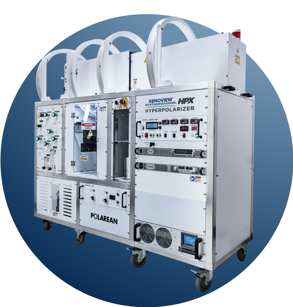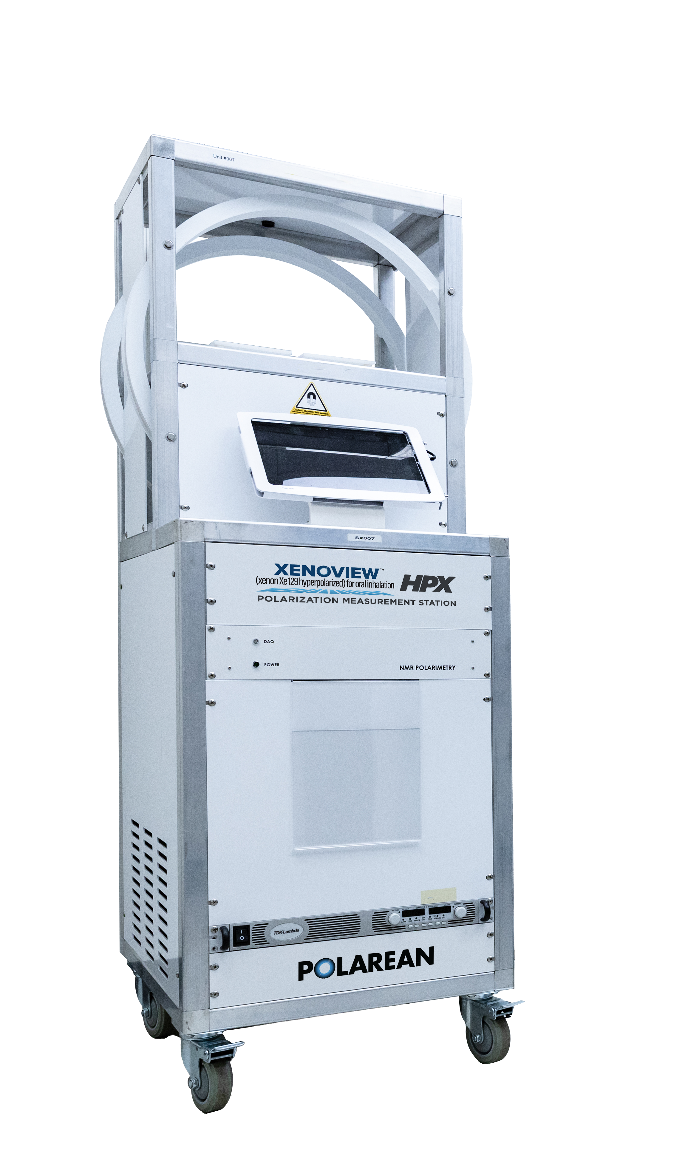
HPX Hyperpolarizer

The HPX Hyperpolarizer measures 67” D x 28” W x 63.25” H.
The HPX Hyperpolarizer processes a Xenon Xe 129 Gas Blend into pure hyperpolarized xenon Xe 129 by using circularly polarized laser light. Hyperpolarized xenon Xe 129 creates a unique MRI signal (resonance frequency) that can be detected to determine its distribution throughout the lungs. Compared to air that is typically within the lung, hyperpolarized xenon Xe 129 enhances the MRI signal by a factor of 100,000.
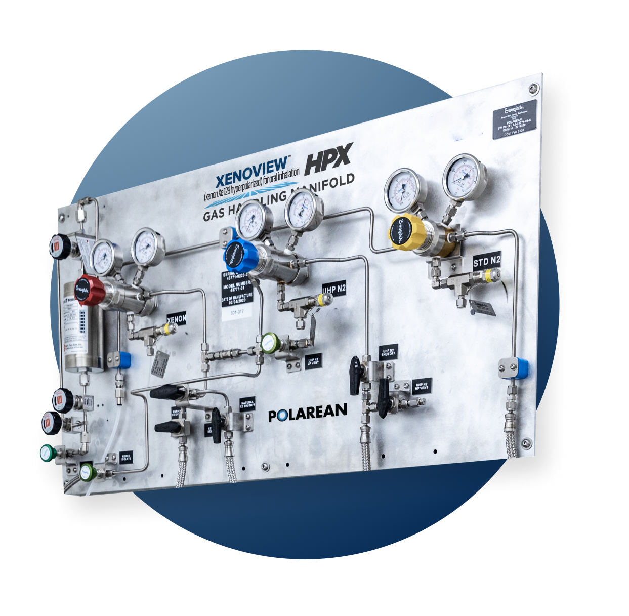
HPX Gas Handling Manifold
The HPX Gas Handling Manifold is an ultra-high purity (UHP) gas pressure regulation and purification system that allows two Xenon Xe 129 Gas Blend cylinders, one NF-grade UHP nitrogen cylinder, and one industrial nitrogen cylinder to be connected to the HPX Hyperpolarizer.
Storage and Requirements

The HPX Gas Handling Manifold measures 48.25″ D x 28.25″ W x 9.25″ H.
Facility Requirements
An MRI scanner is not part of the HPX Hyperpolarization System, but is an essential component of the imaging workflow. The MRI scanner must be an FDA cleared medical device equipped with multi-nuclear (broadband) spectroscopy (MNS) capabilities and have available an appropriate imaging protocol to detect a xenon Xe 129 signal.
The HPX Hyperpolarizer is typically installed in a separate room near the MRI suite for ease of radiology workflow. It measures 67″ L x 28″ W x 63.25″ H and requires a minimum room size of 10′ x 10′, 3-phase electrical power, and compressed air. The room must be sufficiently well ventilated to remove the heat generated by the laser and cell heater.
WORKFLOW
- Compatible with most existing institutional MRI equipment fitted with a multi-channel amplifier
- Programmable Gradient Related Echo (GRE) pulse sequences for xenon Xe 129 MRI scans can be easily integrated into existing radiology workflows
PATIENT EXPERIENCE
- Non-effort dependent procedure
- Short duration in MRI scanner and patient’s head is positioned near opening during procedure
- No radiation exposure
- Inert gas is rapidly exhaled (majority of dose gone within a minute) and no known systemic metabolism or off-organ toxicity
OUTPUT
- Standard DICOM output for standard PACS workstations and/or import into post-processing software programs
- Classified ventilation images, summary reports, and data files containing quantitative statistical analysis.
DICOM, Digital Imaging and Communications in Medicine; PACS, picture archiving and communication system.

